A restoration can take longer than expected even when its design is digital, and a computer-generated file cannot correct a poorly prepared tooth or an unreliable scan. By the end, you will understand each CAD-CAM stage, know when same-day treatment is realistic, and have specific points to compare when discussing digital or conventional treatment.
Key takeaways
- Ask where scanning, design and manufacturing take place.
- Confirm the restoration material matches the tooth and bite demands.
- Check fit at the margins, contacts and opposing teeth.
- Choose digital workflows when they improve verification, not just speed.
What Happens in a CAD-CAM Restoration Workflow
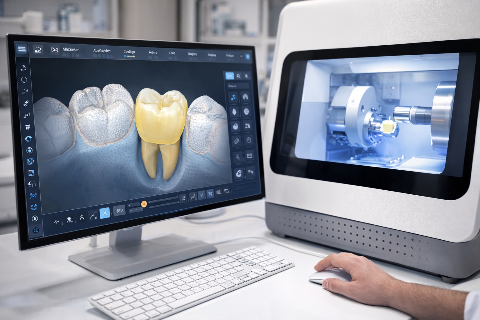
A prepared tooth becomes a finished restoration through a digital chain: CAD-CAM dentistry uses computer-aided design to create the restoration on screen, then computer-aided manufacturing mills or prints it from a selected material. The digital restoration workflow can take place in a dental practice, a laboratory, or both.
1. The dentist examines the tooth and reviews X-rays before removing decay and shaping the preparation. A poor margin, excessive taper, or insufficient reduction remains a problem after scanning.
2. An intraoral scanner records the prepared tooth, nearby teeth, opposing arch, and bite relationship. If scanning is unsuitable, a conventional impression supplies the laboratory with a physical mould.
3. Design software creates the crown, inlay, onlay, veneer, or bridge from those records. The dentist checks the margin, contact points, contour, and proposed bite before approving the design.
4. The software sends the file to a milling machine or 3D printer. The selected ceramic, resin, or other material determines the manufacturing method and the restoration’s required thickness.
5. The restoration receives its material-specific finishing. Zirconia usually requires sintering, while some glass ceramics require crystallization before polishing or glazing.
6. The dentist tries the restoration in the mouth, checks its seating and contacts, and adjusts the bite. A digital proposal cannot replace this clinical check.
7. The restoration is bonded or cemented to the tooth, or attached with a screw in an implant case. Follow-up confirms comfort, hygiene access, and stable function.
How Digital Scans Replace or Supplement Impression Trays
An intraoral scanner captures the visible three-dimensional surfaces of prepared teeth, neighbouring teeth, opposing teeth and the way the arches meet. It records these images as a digital impression, allowing CAD software to design a restoration without a tray filled with impression material.
The scanner records exposed geometry, not tissue hidden beneath it. Moisture, saliva, blood, reflective surfaces and limited access can create gaps or distorted areas, especially around a subgingival margin. Tissue retraction, suction and dry-field isolation still matter; a digital scan does not rescue an unclear preparation boundary.
A conventional impression remains the safer choice when:
- The preparation margin extends below the gum and cannot be exposed clearly.
- Bleeding or persistent saliva prevents a clean scan.
- Limited mouth opening, deep posterior teeth or reflective restorations block scanner access.
- A long-span bridge or full-arch implant case makes stitching many scans together more error-prone.
For single crowns and short-span restorations, an intraoral scanner generally provides accurate working data and reduces physical impression handling. Full-arch scanning demands greater technique control because small alignment errors can accumulate across successive images. A conventional impression also does not remove the need for accurate opposing-arch and bite records.
Check proximal contacts and occlusion in the mouth, because an articulated digital model cannot perfectly reproduce jaw movement.
Why Digital Restorations Can Be Faster Than Conventional Ones
A restoration is fastest when scanning, design and milling happen in the same practice. That removes impression shipping, laboratory queues and several manual fabrication stages, making same-day dentistry practical for many single crowns, inlays, onlays and some veneers.
| Option | What reduces turnaround time | What can add appointments |
|---|---|---|
| Chairside CAD-CAM | Digital files move directly to design and a milled restoration is produced in the practice | Crystallization, sintering, staining, glazing, polishing, bite checks and cementation still take time |
| Laboratory CAD-CAM | Digital transfer avoids a physical impression shipment and speeds laboratory fabrication | The laboratory’s queue, complex design or extensive characterization can require a temporary restoration and a later fitting visit |
| Complex treatment | Digital planning improves coordination between the dentist and laboratory | Bridges, extensive implant restorations and difficult bite or gum conditions often need try-in and staged visits |
A same-day crown is not automatically finished when milling ends. Zirconia usually requires sintering, while some ceramics require crystallization or surface characterization; the dentist must still check the margins, contacts and bite before bonding or cementation.
Expect several appointments when the preparation margin is below the gum, saliva or blood prevents a clean scan, or the tooth needs tissue management. Multiple visits also make sense when:
- The preparation needs correction before fabrication.
- The restoration requires laboratory layering or detailed colour matching.
- An implant case needs surgical healing or a custom abutment.
- The tooth’s nerve condition or structural strength needs monitoring.
A temporary restoration protects the prepared tooth during that interval, but it does not replace diagnosis, isolation or a properly sealed final restoration.
What Precision Means at the Tooth and Bite Level
A restoration fits and functions well only when the tooth preparation, scan and bite record agree. Excessive taper, inadequate occlusal reduction, unsupported ceramic, sharp internal angles or an unclear finish line can produce a weak, bulky or poorly fitting crown despite accurate milling.
| Detail | What the design must achieve | What you need to check |
|---|---|---|
| Marginal fit | Follow the finish line without an open edge or overhang | Inspect the margin clinically; a 120-micrometre gap is a historical benchmark, not a guarantee of success |
| Contact points | Touch adjacent teeth firmly enough to prevent food impaction without blocking seating | Floss should pass with resistance, not tear or stop |
| Occlusal contacts | Meet the opposing teeth evenly without a high spot or damaging load | Check in the patient’s bite and during movement, not only on the digital model |
| Emergence profile | Rise from the gumline in a contour that permits cleaning and supports the soft tissue | Look for overcontouring, pressure on the gum and difficult floss access |
CAD software can suggest proximal contacts and an occlusal scheme, but its result depends on the preparation, opposing-arch scan and bite registration. Digital articulation does not reproduce every jaw movement, so verify contact points and occlusal contacts intraorally.
A good fit cannot rescue a biologically unsound tooth. Confirm isolation, a sealed margin and the correct cementation or bonding protocol; sensitivity or pulpal problems can originate in the tooth rather than the digital workflow.
How to Judge Materials, Verification and Clinical Suitability
A faster workflow is not automatically the better choice: select the material for the tooth’s load, available thickness, optical demands and bonding plan, then verify the result clinically.
| Material or workflow | Main advantage | What to verify |
|---|---|---|
| Lithium disilicate | Natural translucency and reliable bonding for suitable crowns and veneers | Adequate thickness, crystallization, margin fit, shade and occlusion |
| Zirconia | High strength; monolithic designs avoid a separate porcelain veneer and reduce chipping risk | Grade and translucency, thickness, sintering, surface finishing and cementation instructions |
| Printed provisional or guide | Useful for temporary restorations, models and selected implant procedures | Resin indication, post-processing, accuracy, wear, aging and validated use |
A digital plan remains a proposal until the mouth confirms it. Check contacts, margins, seating, bite, shade and cleanability at try-in; a historical 120-micrometre marginal-gap benchmark does not predict success by itself. For implants, compare the planned position with the achieved position because scan-registration error, guide movement, drill tolerance and surgical technique can create deviation.
At Sri Krishna Multispecialty Dental & Implant Studio, ask how the team will verify the scan, design, material processing and final fit rather than choosing “digital” as if it were a material.
Also ask how files are stored and exported; calibration, software compatibility and secure handling determine whether another clinician or laboratory can use the record.
Related services
Crowns & Bridges Precision-Fit Restorations for Damaged or Missing Teeth – Designed Digitally for Function and Esthetics Crowns and bridges are fixed dental... View service → |
Child Dental Health Pediatric Dentistry- Gentle, Specialized Dental Care for Infants, Children, and Teens Pediatric Dentistry Gentle, Specialized Dental Care for Infants, Children, and Teens Children require a special approach to dental care—one that is... View service → |
Frequently asked questions
What happens during a CAD-CAM restoration workflow?
The dentist prepares the tooth, captures digital data, designs the restoration on a computer, manufactures it by milling or printing, then checks its fit and bite before placement.
Do digital scans replace impression trays?
An intraoral scanner can capture the prepared tooth and surrounding teeth without impression material. Dentists may still use conventional impressions when scanning access, moisture control or case complexity makes them more reliable.
Why can CAD-CAM restorations be faster?
A practice with in-house scanning, design and manufacturing can reduce laboratory shipping and appointment delays. Cases sent to an external laboratory still depend on laboratory production and delivery times.
What does precision mean in a CAD-CAM restoration?
Precision includes the restoration’s marginal fit, contact with adjacent teeth, internal seating and relationship with the opposing teeth during biting and movement.
How should you judge a CAD-CAM restoration?
Ask which material is being used, where it is manufactured, how the fit and bite will be verified, and whether the material suits the tooth’s location, function and clinical condition.